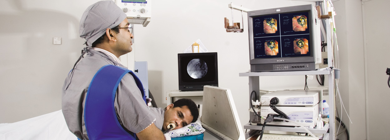
Stone extraction
CBD & CHD moderately dilated, there were multiple small sized smooth surfaced stones, hard in consistency. We did a safe limited papillotomy, and could comfortably extract all the stones by extractor balloon sweeping. The patient was very happy.
Stone extractionPosted by Crescent Gastroliver & General Hospital on Sunday, October 18, 2015
Limited Papillotomy
Bile duct was dilated, and there were multiple stones, small sized. We did a limited papillotomy, and could comfortably extract all the stones. It was rather very safe papillotomy.
Limited PapillotomyPosted by Crescent Gastroliver & General Hospital on Sunday, October 18, 2015
Bleeding during Precut papillotomy
While doing ERCP for Stone extraction, cannulation was difficult, so we attempted pre-cut needle papillotomy. Bleeding occurred, not significantly threatening, we successfully cauterised with the same needle diathermy (ERBE). Haemostasis was confirmed noticing no bleeding even after the stone extraction.
Bleeding during Precut papillotomyPosted by Crescent Gastroliver & General Hospital on Thursday, October 8, 2015
Metallic re-Stenting
It is a very interesting case of metallic (SEMS) re-stenting. In February 2014, ERCP+ metallic stenting (covered) was done; then the diagnosis was Ampullary Growth. Histology finding was negative for malignancy then. He was otherwise alright since 1 month back, but in between this time he was diagnosed as a case of Ca-rectum; and for which he received 25 radiotherapy. He now developed obstructive jaundice for last 1 month. We assumed that the previous stent might have been blocked. We did an ERCP today. Astonishingly we could not find the previous metallic Stent. The covered SEMS expelled out spontaneously and without the notice of the patient. Growth didn�t seem to be aggravated, but bled on touch. CBD was packed with sludge ball with narrowing at lower end. This time we put an uncovered metallic Stent, established free flow of bile. We took a biopsy again (histology awaited).
Metallic re-StentingPosted by Crescent Gastroliver & General Hospital on Thursday, September 17, 2015
Pancreatic Stenting
This patient came from Cox'sbazar, he was suffering pancreatitis for 5 yrs, admitted in hospital 9 times; and then referred to us for ERCP. We found dilated MPD prominently in body and tail with stricture in head region, no definite stone (may be inerstitial/2nd gen duct?). Biliary tree not dilated. It was not easy to cannulate pancreatic duct, we did it without going into CBD, successfully did selective pancreatic papillotomy, then put a pancreatic stent 7 Fr 7 cm in MPD across & above the stricture, free flow of pancreatic secretion noticed.
Pancreatic StentingPosted by Crescent Gastroliver & General Hospital on Wednesday, September 16, 2015
Bleeding after Precut during ERCP
Sometimes, during ERCP, minor bleeding may occur, higher risk in precut needle papillotomy than conventional sphincterotomy. In most of the cases it could be easily manageable. Here we used ERBE endocut diathermy to coagulate, and bleeding stopped before further manipulation.
Bleeding after Precut during ERCPPosted by Crescent Gastroliver & General Hospital on Wednesday, August 19, 2015
Choledocholithiasis with Peri-ampullary diverticulum
There is a big diverticulum. It was really very tricky to do a careful precut with niddle knife, which was extended further by conventional papillotome; and then a very big stone could be extracted with balloon sweeping.
Choledocholithiasis with Peri-ampullary diverticulumPosted by Crescent Gastroliver & General Hospital on Saturday, August 1, 2015
ERCP: Stone Extraction
The papilla was very small and it was just below the mucosal fold, we did very limited papillotomy. Alhamdulillah, through that we could extract the moderate size stone by balloon sweeping, no complication occurred.
ERCP: Stone ExtractionPosted by Crescent Gastroliver & General Hospital on Saturday, August 8, 2015
Worm extraction (Fishing?)
It is a very interesting VDO of worm extraction. The manipulation is like fishing. We awaited outside the papillary opening with the bait for fish/ with the open dormia basket, the ascaris came out ? went inside the trap, and we grasped the worm, then took it out.
Worm extraction (Fishing?)Posted by Crescent Gastroliver & General Hospital on Wednesday, August 19, 2015
ERCP-Huge Stone
It was also a difficult papilla. Biliary tree was grossly dilated, containing multiple filling defects. When we went to extract stone, huge thick pus, biliary sludge and huge number of stones of different sizes, shapes and different colors (some are black, some yellow, spotted) came out. We had to extend papillotomy to evacuate completely. Bleeding occurred, alhamdulillah we managed it.
ERCP-Huge StonePosted by Crescent Gastroliver & General Hospital on Friday, June 19, 2015
Stone Extraction
The papilla was bulged out, luckily it was impacted stone only. We did careful limited sphincterotomy, still it bled, haemostasis was done, the big impacted stone could be extracted easily with balloon.
Stone ExtractionPosted by Crescent Gastroliver & General Hospital on Monday, July 27, 2015
Stone extraction
Small papilla, wasn't difficult to enter, but required limited papillotomy + sphincteroplasty, could easily take out the moderately big sized stone with extractor balloon.
Stone extractionPosted by Crescent Gastroliver & General Hospital on Saturday, July 25, 2015
Worm Extraction
It is never easy to grasp a worm/AL inside CBD & take it out, Specially when the pt is gravid, 12 weeks or 29 wks, restricting visualisation in Xray fluoro. We folded it on itself inside CBD and grasped it with dormia basket, & extracted it with minimum radiation (though mother pt is securely shielded on uterus)... that is the technique
Worm ExtractionPosted by Crescent Gastroliver & General Hospital on Wednesday, July 29, 2015
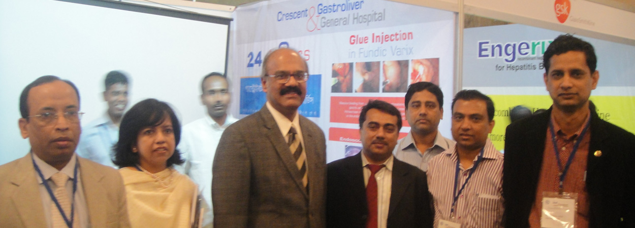
Glue Inj in Fundic Varix
This lady came with 4 episodes of severe hematemesis & malena in last few yrs. Each times she vomitted for 7-8 times/per day, more than 100-200 cc apprx in each vomit, lowering Hb% to 7gm around. She was diagnosed as a case of NBNC CLD (we r thinking of wilson's). This time when she reached us from Sylhet, we started Inj. Stilamin, & tried to settle her with blood transfusion ..... We repeated Endoscopy, which showed large Oes Varices, and Fundic Varices with evidence of recent bleeding. We injected Cyanoacrylate Glue in the big varix targeting near the point of bleeding. It is a Unique procedure done in our set up; most of the centres don't show interest as improper technique or slight breech of caution may damage a costly Endoscope. We r alhamdulillah doing it for yrs safely.
Glue Inj in Fundic VarixPosted by Crescent Gastroliver & General Hospital on Tuesday, May 26, 2015
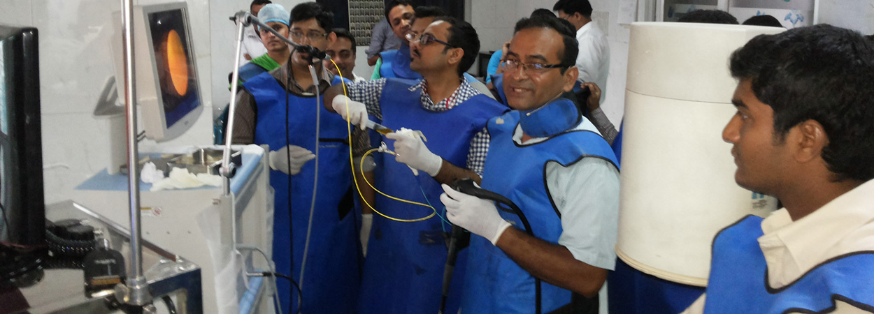
ERCP + Stone extraction
Pt reported for repeat ERCP (recurrent Cholodocholithiasis). Papilla was not easy to find out and aproach. But we succeeded, stone was soft though big.
ERCP + Stone extractionPosted by Crescent Gastroliver & General Hospital on Friday, June 5, 2015
Precut Papillotomy
Atypical growth at ampulla of vater, ?malignant. Moribund patient with IHD/EF-only 45%, Position at duodenum was not comfortable, we had to go for precut papillotomy with niddle-knife. It was a splendid precut, exploring the feshy growth inside, biopsy was taken, histology awaited. We quickly put a stent. Indeed a very smart safe anesthesia, thnx Dr Alamgir Haider.
Precut PapillotomyPosted by Crescent Gastroliver & General Hospital on Monday, May 18, 2015
ERCP-Stenting for post-Op Biliary leakage
This is a unique case. She was operated on - Cholecystectomy+ Choledocholithotomy (huge number stones were retrieved). T-tube was placed, upto 15 POD was uneventful. Clamping T-tube started on POD-13, and finally removed on 15. Then there was evidence for biliary leakage, relaparotomy could not find the point of leakage/?injury. 2 drain was placed and she was referred to us. With biliary peritonitis, her Hb-8gm, Alb-2.8 gm/dl, Serum K+ 2.9 mmol/L, and bil pleural effusion, she could not maintain Oxygen saturation satisfactorily. We did ERCP, didn't try to extract the stones, but put a stent across & above the stones and point of leakage (from the level where T-tube was inserted). Pt started improving.
ERCP-Stenting for post-Op Biliary leakagePosted by Crescent Gastroliver & General Hospital on Monday, May 18, 2015
ERCP + Stone extraction
It was really a very difficult ERCP; bilateral diverticulum made cannulation, as well as papillotomy difficult and limited, stone was very big, there were possibly stricture in some part of biliary tree with proximal dilatation. However stone was soft and we could clear the tree, to benefit the patient.
ERCP + Stone extractionPosted by Crescent Gastroliver & General Hospital on Sunday, May 17, 2015

 A Sister Concern of